Picture a physicist. Do you imagine someone madly scribbling equations about nuclear energy or black holes? What about someone doing research on dementia? Or figuring out better ways to do high-tech medical imaging? These are the sorts of things a Magnetic Resonance Imaging (MRI) physicist does.
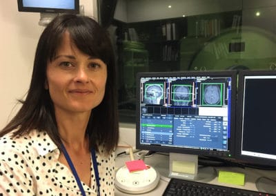
Catherine Morgan is one of only a handful of MRI physicists across New Zealand. She has a split job: half her time, she’s a research fellow at the University of Auckland’s School of Psychology, while the other half of the time, she’s the senior MRI physicist at the Centre for Advanced MRI (CAMRI), a UniServices business unit based at the Faculty of Medical and Health Sciences that offers cutting-edge imaging for both clinical and research purposes.
In some respects, the two aspects of Morgan’s job are quite different – but both her research and clinical work are solidly focused on helping people.
Morgan’s path to what she’s doing now was a bit circuitous. As a young girl growing up in England, she enjoyed physics, so when she went to university, that’s what she majored in. When she graduated, she got a job as a “quant” – a quantitative analyst who does financial modelling – at a property company in London.
“I did that for about a year, then I found myself in a meeting at two o’clock in the morning about tax and accounting codes for a bid we were putting in the next day and I just thought, ‘This is not for me or what I want to do with my life,’” says Morgan.
It was Morgan’s mother, a nurse, who suggested medical physics. Taking advantage of a National Health Service (NHS) training scheme, Morgan enrolled in a master’s programme that combined working in hospitals with part-time study. She loved it and went on to do a PhD in the field.
“I really enjoy my job. It doesn’t feel like work,” says Morgan. “I’d be richer if I’d stuck with tax and accounting, but this is a real opportunity for me to do something meaningful as a physicist.”
The physics of MRI
MRI has a lot of complex physics behind it. Invented in the 1970s, it relies on powerful magnets that produce such a strong, uniform magnetic field that it forces most of the water molecules in your body – or more specifically, the hydrogen protons in them – to line up.
An MRI scan directs radio frequency pulses at specific parts of the body, forcing the water molecules at a lower energy state to spin around. When the pulse stops, they return to their original state and release the energy they temporarily absorbed. Because different types of tissue release energy differently, MRI can use this to create detailed images of what’s inside our bodies.
By changing settings such as the timing and type of radio frequency or by making small adjustments to the magnetic field, researchers and clinicians can focus on different elements even within the same part of the body. For example, one brain scan might focus on white matter while another might measure blood flow.
“You can either use settings that come from the manufacturer of the scanner or, if you have a knowledge of how it works, you can change those settings,” says Morgan. “That’s where I often come in. New ways of doing things can lead to better-informed, clearer diagnoses for clinical work or new discoveries on the research side. For example, there’s lots of work being done in areas such as how our brain ages or how visual processing works. It’s helping us understand normal brain functioning, which is still a bit mysterious.”
As CAMRI’s MRI physicist, Morgan works with medical imaging technologists to help researchers and clinicians with new and better ways of doing imaging. One week she might be checking the quality of new advanced methods for imaging prostate cancer, while the next week, she might be matching CAMRI scanner settings to another centre in Canada for a joint project on whether preterm babies have different outcomes based on their nutrition. She regularly talks to MRI physicists around the world about new types of scans for hearts, brains and more.
“I really enjoy how varied the work is,” says Morgan.
Finding early predictors of dementia
Morgan’s own research focuses on brain imaging to understand how dementia develops. She’s part of a large national study, the Dementia Prevention Research Clinics (DPRCs), studying people with mild cognitive impairment (MCI) in Aotearoa. People with MCI have marked deficits in cognition but can cope with everyday tasks and live independently. Some people deteriorate into dementia while others do not.
The DPRC team is following more than 250 participants nationally over years, regularly taking MRI scans, blood samples, medical assessments and tests examining multiple domains of cognition. The project aims to find early signs that differentiate people who progress to Alzheimer’s disease and those who don’t.
From the MRI scans, researchers will examine factors such as brain volume, since brains shrink in dementia, as well as blood flow in the brain and white matter tracts, which connect different regions of the brain. The scan results, when combined with other data from the study, have the potential to be groundbreaking, but the MRI techniques themselves are well established.
Morgan is leading a smaller side study using brand new MRI methods. One scanning method focuses on iron deposition, since some evidence suggests elevated brain iron levels can be damaging. Another examines blood flow in the brain using a new type of scan originally developed for the heart, known as 4D flow, which allows for precise measurements of blood flow through a vessel.
A third new technique examines the blood-brain barrier, a protective layer around the blood vessels in the brain. New evidence suggests the blood-brain barrier may be dysfunctional in MCI. Normally a contrast agent – a chemical injected into a patient to improve clarity of images – would be used. However, there is some concern that some patients who have many MRIs using certain forms of the contrast agent could end up with small amounts of gadolinium, a metal used in the contrast agent, building up in the brain and other tissues. That’s why Morgan and her team are trying a new technique focusing on water molecules already in our bodies.
“We may not need to use contrast agents at all. Or we may uncover a new aspect of brain health related to water transport. It’s an exciting study and we’re close to putting it all together,” says Morgan. “If we can uncover something people haven’t thought of before in terms of the way Alzheimer’s develops, there could be new options for treatments.”
If you have a query about how advanced MRI techniques could apply to your research or enhance your imaging outcomes, contact Catherine Morgan.
– This article first appeared on UniServices